It has the action of a force pump
As the blood is forced through the heart by forcible contractions of its muscular walls, it has the action of a force pump, and gives the impulse at each beat, which we call the pulse the dilatation of the arteries throughout the system. The contraction of the auricles is quickly followed by that of the ventricles, and then a slight pause occurs; this takes place in regular rhythmical order during health.
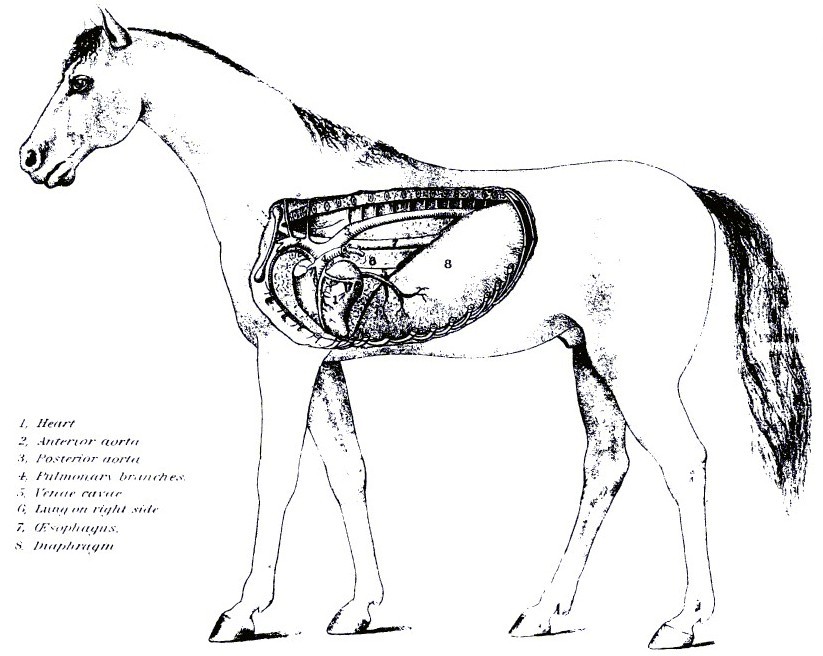
PLATE XX.
INTERIOR OF CHEST SHOWING POSITION OF HEART AND DIAPHRAGM.

PLATE XXI.
CIRCULATORY APPARATUS.
The action of the heart is governed and maintained by the pneumogastric nerve (tenth pair of cranial nerves); it is the inhibitory nerve of the heart, and regulates, slows, and governs its action. When the nerve is cut, the heartbeats increase rapidly, and, in fact, the organ works without control. When the nerve is unduly irritated the holdback, or inhibitory force, is increased, and the heart slows up in the same measure. The left cavities of the heart, the pulmonary veins, and the aorta, or systemic artery, contain red or florid blood, fit to circulate through the body. The right cavities of the heart, with the venae cavae, or systemic veins, the pulmonary artery, contain dark blood, which must be transmitted through the lungs for renovation.
The arteries, commencing in two great trunks, the aorta and the pulmonary artery, undergo division, as in the branching of a tree. Their branches mostly come off at acute angles, and are commonly of uniform diameter in each case, but successively diminish after and in consequence of division, and in this manner gradually merge into the capillary system of blood vessels. As a general rule, the combined area of the branches is greater than that of the vessels from which they emanate, and hence the collective capacity of the arterial system is greatest at the capillary vessels. The same rule applies to the veins. The effect of the division of the arteries is to make the blood move more slowly along their branches to the capillary vessels, and the effect of the union of the branches of the veins is to accelerate the speed of the blood as it returns from the capillary vessels to the venous trunks.
In the smaller vessels a frequent running together, or anastomosis, occurs. This admits of a free communication between the currents of blood, and must tend to promote equability of distribution and of pressure, and to obviate the effects of local interruption. The arteries are highly elastic, being extensile and retractile both in length and breadth. During life they are also contractile, being provided with muscular tissue. When cut across they present, although empty, an open orifice; the veins, on the other hand, collapse.
In most parts of the body the arteries are inclosed in a sheath formed of connective tissue, but are connected so loosely that, when the vessel is cut across, its ends readily retract some distance within the sheath. Independently of this sheath, arteries are usually described as being formed of three coats, named, from the relative positions, external, middle, and internal. This applies to their structure so far as it is discernible by the naked eye. The internal, serous, or tunica intima, is the thinnest, and is continuous with the lining membrane of the heart. It is made up of two layers an inner, consisting of a layer of epithelial scales, and an outer, transparent, whitish, highly elastic, and perforated. The middle coat, tunica media, is elastic, dense, and of a yellow color, consisting of nonstriated muscular and elastic fibers, thickest in the largest arteries and becoming thinner in the smaller. In the smallest vessels it is almost entirely muscular. The external coat, tunica adventitia, is composed mainly of fine and closely woven bundles of white connective tissue, which chiefly run diagonally or obliquely around the vessel. In this coat the nutrient vessels, the vasa vasorum, form a capillary network, from which a few penetrate as far as the muscular coat.
The veins differ from arteries in possessing thinner walls, less elastic and muscular tissue, and for the most part a stronger tunica adventitia. They collapse when cut across or when they are empty. The majority of veins are provided with valves; these are folds of the lining membrane, strengthened by fibrous tissue. They favor the course of the blood and prevent its reflux. The nerves which supply both the arteries and the veins come from the sympathetic system. The smaller arteries terminate in the system of minute vessels known as the capillaries, which are interposed between the termination of the arteries and the commencement of the veins. Their average diameter is about one three-thousandth of an inch.
Nenhum comentário:
Postar um comentário