quinta-feira, 31 de dezembro de 2015

Spinal Congestion.

Spinal Congestion.

Spinal Congestion.


This condition consists in an excess of blood. As the blood vessels of the pia mater are the principal source of supply to the spinal cord, hyperemia of the cord and of the meninges usually go together. The symptoms are, therefore, closely allied to those of spinal meningitis and congestion. When the pia mater is diseased, the spinal cord is almost invariably affected also.

Cause. Sudden checking of the perspiration, violent exercise, blows, and falls.

quarta-feira, 30 de dezembro de 2015

The inflammation may involve nearly the whole length of the cord

The inflammation may involve nearly the whole length of the cord

The inflammation may involve nearly the whole length of the cord


Pathology. The inflammation may involve nearly the whole length of the cord, but generally it is more intense in some places than others; when due to mechanical injury, the inflammation may remain confined to a small section. The cord is swollen and congested, reddened, often softened and infiltrated with pus cells, and the nerve elements are degenerated.

Treatment. Similar to that of spinal meningitis.

terça-feira, 29 de dezembro de 2015

The attack may begin with a chill or convulsion

The attack may begin with a chill or convulsion

The attack may begin with a chill or convulsion


Symptoms. The attack may begin with a chill or convulsion; the muscles twitch or become cramped very early in the disease, and the bladder usually is affected at the outset, in which there may be either retention or incontinence of urine. These conditions are followed by complete or partial paralysis of the muscles posterior to the locality of the inflamed cord, and the muscles begin to waste away rapidly. The paralyzed limb becomes cold and dry, due to the suspension of proper circulation; the joints may swell and become edematous; vesicular eruptions appear on the skin; and frequently gangrenous sloughs form on the paralyzed parts. It is exceedingly seldom that recovery takes place. In a few instances it may assume a chronic type, when all the symptoms become mitigated, and thus continue for some time, until septicemia, pyemia, or exhaustion causes death.

segunda-feira, 28 de dezembro de 2015

Myelitis, or Inflammation of The Substance of The Spinal Cord.

Myelitis, or Inflammation of The Substance of The Spinal Cord.

Myelitis, or Inflammation of The Substance of The Spinal Cord.


This is a rare disease, except as a secondary result of spinal meningitis or injuries to the spine. Poisoning by lead, arsenic, mercury, phosphorus, carbonic-acid gas, etc., has been known to produce it. Myelitis may be confined to a small spot in the cord or may involve the whole for a variable distance. It may lead to softening abscess or degeneration.

domingo, 27 de dezembro de 2015

Bags filled with ice should be applied along the spine

Bags filled with ice should be applied along the spine

Bags filled with ice should be applied along the spine


Treatment. Bags filled with ice should be applied along the spine, to be followed later by strong blisters. The fever should be controlled as early as possible by giving 20 drops of Norwood's tincture of veratrum viride every hour until the desired result is obtained. One dram of the fluid extract of belladonna, to control pain and vascular excitement of the spinal cord, may be given every five or six hours until the pupils of the eyes become pretty well dilated. If the pain is very intense 5 grains of sulphate of morphia should be injected hypodermically. The animal must be kept as free from excitement as possible. If the urine is retained in the bladder it must be drawn off every four or six hours. In very acute attacks the disease generally proves fatal in a few days. If, however, the animal grows better, some form of paralysis is liable to remain for a long time, and the treatment will have to be directed then toward a removal of the exudative products and a strengthening of the system and stimulation of the nervous functions. To induce absorption, iodid of potassium in 2-dram doses, dissolved in the drinking water, may be given twice a day. To strengthen the system, iodid of iron 1 dram twice a day and 1 dram of nux vomica once a day may be given in the feed. Electricity to the paralyzed and weakened muscles is advisable; the current should be weak, but be continued for half an hour two or three times daily. If the disease is due to a broken back, caries of the vertebrae, or some other irremediable cause, the animal should be destroyed at once.

sábado, 26 de dezembro de 2015

There is an effusion of serum between the membranes

There is an effusion of serum between the membranes

There is an effusion of serum between the membranes


Pathology. In spinal meningitis we find essentially the same condition as in cerebral meningitis; there is an effusion of serum between the membranes, and often a plastic exudation firmly adherent to the pia mater serves to maintain a state of paralysis for a long time after the acute symptoms have disappeared by compressing the cord. Finally, atrophy, softening, and even abscess may develop within the cord. Unlike in man, it is usually found localized in horses.

sexta-feira, 25 de dezembro de 2015

Spinal Meningitis, or Inflammation of The Membranes Enveloping The Spinal Cord.

Spinal Meningitis, or Inflammation of The Membranes Enveloping The Spinal Cord.

Spinal Meningitis, or Inflammation of The Membranes Enveloping The Spinal Cord.


This may be induced by the irritant properties of blood poisons, exhaustion and exposure, spinal concussion, all forms of injury to the spine, tumors, caries of the vertebrae, rheumatism, etc.

Symptoms. A chill may be the precursor, a rise in temperature, or a general weakness and shifting of the legs. Soon a painful, convulsive twitching of the muscles sets in, followed by muscular rigidity along the spine, in which condition the animal will move very stiffly and evince great pain in turning. Evidences of paralysis or paraplegia develop, retention or incontinence of urine, and oftentimes sexual excitement is present. The presence of marked fever at the beginning of the attack, associated with spinal symptoms, should lead us to suspect spinal meningitis or myelitis. These two conditions usually appear together, or myelitis follows inflammation of the meninges so closely that it is almost impossible to separate the two; practically it does not matter much, for the treatment will be about the same in both cases. Spinal meningitis generally becomes chronic, and is then marked principally by paralysis of that portion, or parts of it, posterior to the seat of the disease.

quinta-feira, 24 de dezembro de 2015

In all paralytic affections there may be anesthesia

In all paralytic affections there may be anesthesia

In all paralytic affections there may be anesthesia


Treatment for paralysis. In all paralytic affections there may be anesthesia, or impairment of sensibility, in addition to the loss of motion, or there may be hyperesthesia, or increased sensibility, in connection with the loss of motion. These conditions may call for special treatment in addition to that for loss of motion. If hyperesthesia is well marked local anodynes may be needed to relieve suffering. Chloroform liniment or hypodermic injections of from 3 to 5 grains of sulphate of morphia will allay local pain. If there is marked anesthesia, or loss of sensibility, it may become necessary to secure the animal in such way that he can not suffer serious injury from accidents which he can not avoid or feel. In the treatment of any form of paralysis we must always refer to the cause, and attempt its removal if it can be discovered. In cases in which the cause can not be determined we have to rely solely upon a general external and internal treatment. Externally, fly blisters or strong, irritant liniments may be applied to the paralyzed parts. In hemiplegia they should be applied along the bony part of the side of the neck; in paraplegia, across the loins. In, some cases hot-water cloths will be beneficial. Internally, it is well to administer 1 dram of powdered nux vomica or 2 grains of sulphate of strychnia twice a day until twitching of some of the voluntary muscles occurs; then discontinue it for several days, and then commence again with a smaller dose, gradually increasing it until twitching recurs. Iodid of potassium in 1 to 2 dram doses two or three times daily may be used with the hope that it will favor the absorption of the clot or obstruction to the nervous current. In some cases Fowler's solution of arsenic in teaspoonful doses twice a day in the drinking water proves beneficial. Occasionally benefit may be derived from the application of the electric current, especially in cases of roaring, facial paralysis, paralysis of the eyelid, etc. Nutritious but not too bulky feed, good ventilation, clean stabling, moderate exercise if the animal is capable of taking it, good grooming, etc., should be observed in all cases.

quarta-feira, 23 de dezembro de 2015

Partial paralysis of the heart and organs of respiration

Partial paralysis of the heart and organs of respiration

Partial paralysis of the heart and organs of respiration


Paralysis of hearing, of the external ear, of the eyelid, partial paralysis of the heart and organs of respiration, of the blood vessels from injury to the vasomotor nerves of the esophagus, or loss of deglutition, palsy of the stomach, all may be manifested when the supply of nervous influence is impaired or suspended.

terça-feira, 22 de dezembro de 2015

This usually affects the neck of the bladder

This usually affects the neck of the bladder

This usually affects the neck of the bladder


Paralysis of the bladder. This usually affects the neck of the bladder, and is characterized by incontinence of urine; the urine dribbles away as fast as it is secreted. The cause may be of reflex origin, disease of the rectum, tumors growing within the pelvic cavity, injury to the spinal cord, etc.

Paralysis of the optic nerve (amaurosis). A paralysis of eyesight may occur very suddenly from rupture of a blood vessel in the brain, acute local congestion of the brain, the administration of excessive doses of belladonna or its alkaloid atropia, etc. In amaurosis the pupil is dilated to its full extent; the eye looks clear, but does not respond to light.

segunda-feira, 21 de dezembro de 2015

Paralysis of the rectum and tail

Paralysis of the rectum and tail

Paralysis of the rectum and tail


Paralysis of the rectum and tail. This is generally the result of a blow or fall on the rump, which causes a fracture of the sacrum bone and injury to the nerves supplying the tail and part of the rectum and muscles belonging thereto. This fracture would not be suspected were it not for the loss of motion of the tail.

Intestinal paralysis. Characterized by persistent constipation; frequently the strongest purgatives have no effect whatever on the movement of the bowels. In the absence of symptoms of indigestion, or special diseases implicating the intestinal canal, torpor of the bowels must be attributed to deficient innervation. This condition may depend upon brain affections or be due to reflex paralysis. Sudden checks of perspiration may induce excessive action of the bowels or paralysis.

domingo, 20 de dezembro de 2015

A flaccid condition of the cheek muscles

A flaccid condition of the cheek muscles

A flaccid condition of the cheek muscles


Symptoms. A flaccid condition of the cheek muscles, pendulous lips, inability to grasp the feed, often a slow and weak movement in chewing, and difficulty and slowness in drinking.

Laryngismus paralyticus, or roaring. This condition is characterized by roaring, and is usually caused by an inflamed or hypertrophied bronchial gland pressing against the left recurrent laryngeal nerve, which interferes with its conducting power. A similar condition is occasionally induced in acute pleurisy, when the recurrent nerve becomes involved in the diseased process or compressed by plastic exudation.

sábado, 19 de dezembro de 2015

This is frequently met with in horses

This is frequently met with in horses

This is frequently met with in horses


Local paralysis. This is frequently met with in horses. It may affect many parts of the body, even vital organs, and it is very frequently overlooked in diagnosis.

Facial paralysis. This is a frequent type of local paralysis, and is due to impairment of function of the motor nerve of the facial muscles, the portio dura. The cause may exist at the base of the brain, compression along its course after it leaves the medulla oblongata, or to a bruise after it spreads out on the great masseter muscle.

sexta-feira, 18 de dezembro de 2015

The animal appears usually perfectly healthy

The animal appears usually perfectly healthy

The animal appears usually perfectly healthy


Locomotor ataxia, or incoordination of movement. This is characterized by an inability to control properly the movement of the limbs. The animal appears usually perfectly healthy, but when he is led out of his stall his legs have a wobbly movement and he will stumble or stagger, especially in turning. When this is confined to the hind parts it may be termed a modified form of paraplegia, but often it may be seen to affect nearly all the voluntary muscles when they are called into play, and must be attributed to some pressure exerted on the base of the brain.

quinta-feira, 17 de dezembro de 2015

When due to mechanical injury of the spinal cord

When due to mechanical injury of the spinal cord

When due to mechanical injury of the spinal cord


Symptoms. When due to mechanical injury of the spinal cord, from a broken back or spinal hemorrhage, it is generally progressive in its character, although it may be sudden. When it is caused by agents in the blood, it may be intermittent or recurrent.

Paraplegia is not difficult to recognize, for it is characterized by a weakness and imperfect control of the hind legs and powerless tail. The urine usually dribbles away as it is formed and the manure is pushed out, ball by ball, without any voluntary effort, or the passages may cease entirety. When paraplegia is complete, large and ill-conditioned sores soon form on the hips and thighs from chafing and bruising, which have a tendency quickly to weaken the animal and necessitate his destruction.

quarta-feira, 16 de dezembro de 2015

Paraplegia transverse paralysis of the hind extremities

Paraplegia transverse paralysis of the hind extremities

Paraplegia transverse paralysis of the hind extremities


Paraplegia (transverse paralysis of the hind extremities). Paralysis of the hind extremities is usually due to some injury or inflammation affecting the spinal cord. (See "Spinal meningitis," p. 232, and "Myelitis," p. 233.) It may also be due to a reflex irritation from disease of peripheral nerves, to spinal irritation or congestion caused by blood poisons, etc.

terça-feira, 15 de dezembro de 2015

In hemiplegia the attack may be very sudden

In hemiplegia the attack may be very sudden

In hemiplegia the attack may be very sudden


Symptoms. In hemiplegia the attack may be very sudden, and the animal fall, powerless to move one side of the body; one side of the lips will be relaxed; the tongue may hang out on one side of the mouth; the tail curved around sideways; an inability to swallow feed or water may be present, and often the urine dribbles away as fast as it collects in the bladder. Sensibility of the affected side may be entirely lost or only partial; the limbs may be cold and sometimes unnaturally warm. In cases wherein the attack is not so severe the animal may be able to maintain the standing position, but will have great difficulty in moving the affected side. In such cases the animal may recover from the disability. In the more severe, in which there is complete loss of power of movement, recoveries are rare.

segunda-feira, 14 de dezembro de 2015

Hemiplegia paralysis of one side or half of the body

Hemiplegia paralysis of one side or half of the body

Hemiplegia paralysis of one side or half of the body


Hemiplegia (paralysis of one side or half of the body). Hemiplegia is frequently the result of a tumor in the lateral ventricles of the brain, softening of one hemisphere of the cerebrum, pressure from extravasated blood, fracture of the cranium, or it may be due to poisons in the blood or to reflex origin. When hemiplegia is due to or the result of a prior disease of the brain, especially of an inflammatory character, it is seldom complete; it may affect only one limb and one side of the head, neck, or muscles along the back, and may pass off in a few days after the disappearance of all the other evidences of the primary affection. In most cases, however, hemiplegia arises from emboli obstructing one or more blood vessels of the brain, or the rupture of some vessel the wall of which had become weakened by degeneration and the extravasation of blood. Sensibility in most cases is not impaired, but in some there is a loss of sensibility as well as of motion. In some cases the bladder and rectum are involved in the paralysis.

domingo, 13 de dezembro de 2015

This can not take place without producing immediate death

This can not take place without producing immediate death

This can not take place without producing immediate death


General paralysis. This can not take place without producing immediate death. The term is, however, usually applied to paralysis of the four extremities, whether any other portions of the body are involved or not. This form of palsy is due to compression of the brain by congestion of its vessels, large clot formation in apoplexy, concussion, or shock, or any disease in which the whole brain structure is involved in functional disturbance.

sábado, 12 de dezembro de 2015

Disease of the blood vessels of the brain

Disease of the blood vessels of the brain

Disease of the blood vessels of the brain


Causes are much varied. Most of the acute affections of the brain and spinal cord may lead to paralysis. Injuries, tumors, disease of the blood vessels of the brain, etc., all have a tendency to produce suspension of the conducting motive power to the muscular structures. Pressure upon, or the severing of, a nerve causes a paralysis of the parts to which such a nerve is distributed. Apoplexy may be termed a general paralysis, and in nonfatal attacks is a frequent cause of the various forms of palsy.

sexta-feira, 11 de dezembro de 2015

Paralysis, or Palsy.

Paralysis, or Palsy.

Paralysis, or Palsy.


Paralysis is a weakness or cessation of the muscular contraction by diminution of loss of the conducting power or stimulation of the motor nerves. Paralytic affections are of two kinds, the complete and the incomplete. The former includes those in which both motion and sensibility are affected; the latter those in which only one or the other is lost or diminished. Paralysis may be general or partial. The latter is divided into hemiplegia and paraplegia. When only a small portion of the body is affected, as the face, a limb, the tail, it is designated by the term local paralysis. When the irritation extends from the periphery of the center it is termed reflex paralysis.

quinta-feira, 10 de dezembro de 2015

Epilepsy, or Falling Fits.

Epilepsy, or Falling Fits.

Epilepsy, or Falling Fits.


The cause of epilepsy is seldom traceable to any special brain lesions. In a few cases it accompanies disease of the pituitary body, which is located in the under surface of the brain. Softening of the brain may give rise to this affection. Attacks may occur only once or twice a year or they may be of frequent recurrence.

Symptoms. No premonitory symptoms precede an epileptic fit. The animal suddenly staggers; the muscles become cramped; the jaws may be spasmodically opened and closed, and the tongue become lacerated between the teeth; the animal foams at the mouth and falls in a spasm. The urine flows involuntarily, and the breathing may be temporarily arrested. The paroxysm soon passes off, and the animal gets on its feet in a few minutes after the return of consciousness.

Treatment. Dashing cold water on the head during the paroxysm. After the recovery 1 dram of oxid of zinc may be given in the feed twice a day for several weeks, or benefit may be derived from the tonic prescribed for chorea.

quarta-feira, 9 de dezembro de 2015

Chorea is often associated with a nervous disposition

Chorea is often associated with a nervous disposition

Chorea is often associated with a nervous disposition


Chorea is often associated with a nervous disposition, and is not so frequent in animals with a sluggish temperament. The involuntary muscular contractions cause no pain, and do not appear to produce much exhaustion of the affected muscles, although the jerking may be regular and persistent whenever the animal is in motion.

Treatment. In a few cases, early in the appearance of this affection, general nerve tonics may be of benefit, viz, iodid of iron, 1 dram; pulverized nux vomica, 1 dram; pulverized scutellaria (skull-cap), 1 ounce. Mix and give in the feed once a day for two weeks. Arsenic in the form of Fowler's solution is often beneficial. If the cause is connected with organic brain lesions, treatment is usually unsuccessful.

terça-feira, 8 de dezembro de 2015

Stringhalt is by some termed chorea

Stringhalt is by some termed chorea

Stringhalt is by some termed chorea


Stringhalt is by some termed "chorea." This is manifested by a sudden jerking up of one or both hind legs when the animal is walking. This symptom may be very slight in some horses, but has a tendency to increase with age. In some the catching up of the affected leg is very violent, and when it is lowered to the ground the motion is equally sudden and forcible, striking the foot to the ground like a pile driver. Very rarely chorea may be found to affect one of the fore legs, or the muscles of one side of the neck or the upper part of the neck. Involuntary jerking of the muscles of the hip or thigh is seen occasionally, and is termed "shivering" by horsemen.

segunda-feira, 7 de dezembro de 2015

Chorea, or St. Vitus's Dance.

Chorea, or St. Vitus's Dance.

Chorea, or St. Vitus's Dance.


Chorea is characterized by involuntary contractions of voluntary muscles. This disease is an obscure disorder, which may be from pressure upon a nerve, cerebral or spinal sclerosis, small aneurisms in the brain, etc. Choreic symptoms have been produced by injecting granules of starch into the arteries entering the brain. Epilepsy and other forms of convulsions simulate chorea in appearance.

domingo, 6 de dezembro de 2015

Irregular muscular contractions of various parts of the body

Irregular muscular contractions of various parts of the body

Irregular muscular contractions of various parts of the body


Convulsions. Although there is no disease of the nervous system which can be properly termed convulsive, or justify the use of the word convulsion to indicate any particular disease, yet it is often such a prominent symptom that a few words may not be out of place. General, irregular muscular contractions of various parts of the body, with unconsciousness, characterize what we regard as convulsions, and like ordinary spasms are dependent upon some disease or irritation of the nervous structures, chiefly of the brain. No treatment is required; in fact, a general convulsion must necessarily be self-limited in its duration. Suspending, as it does, respiratory movements, checking the oxygenation and decarbonization of the blood, the rapid accumulation of carbonic-acid gas in the blood and the exclusion of oxygen quickly puts the blood in a condition to produce the most reliable and speedy sedative effect upon the nerve excitability that could be found, and consequently furnishes its own remedy so far as the continuance of the convulsive paroxysm is concerned. Whatever treatment is instituted must be directed toward a removal of the cause of the convulsive paroxysm.

sábado, 5 de dezembro de 2015

Composed of chloroform 1 part and soap liniment 4 parts

Composed of chloroform 1 part and soap liniment 4 parts

Composed of chloroform 1 part and soap liniment 4 parts


Treatment of spasms. An anodyne liniment, composed of chloroform 1 part and soap liniment 4 parts, applied to cramped muscles will usually cause relaxation. This may be used when single external muscles are affected. In spasms of the glottis, inhalation of sulphuric ether will give quick relief. In spasm of the diaphragm, rest and the administration of half an ounce of chloroform in 3 ounces of whisky, with a pint of water added, will generally suffice to bring relief, or if this fails give 5 grains of sulphate of morphia by hypodermic injection. If spasms result from organic disease of the nervous system, the latter should receive such treatment as its character demands. In cramp of the leg, compulsory movement usually causes relaxation very quickly; therefore the animal should be led out of the stable and be forced to run or trot. Sudden, nervous excitement caused by a crack of the whip or smart blow will often bring about immediate relief. Should this fail, the anodyne liniment may be used along the inside of the thigh, and chloroform, ether, or laudanum given internally. An ounce of the chloral hydrate will certainly relieve the spasm when given internally, but the cramp may return soon after the effect has passed off, which in many cases it does very quickly.

sexta-feira, 4 de dezembro de 2015

It is probably due to an irritation of the nerves of the thigh

It is probably due to an irritation of the nerves of the thigh

It is probably due to an irritation of the nerves of the thigh


Spasm of the thigh, or cramp of a hind limb. This is frequently witnessed in horses that stand on sloping plank floors generally in cold weather or it may come on soon after severe exercise. It is probably due to an irritation of the nerves of the thigh. In cramps of the hind leg the limb becomes perfectly rigid, and attempts to flex are unsuccessful; the animal stands on the affected limb, but is unable to move it; it is unnaturally cold; it does not, however, appear to cause much suffering unless attempts are made to change position. This cramp may be of short duration a few minutes or it may persist for several days. This condition is often taken for a dislocation of the stifle joint. In the latter the foot is extended backward, and the horse is unable to advance it, but drags the limb. An examination of the joint also reveals a change in form. Spasms may affect the eyelids, by closure or by retraction. Spasm of the sterno-maxillaris muscle has been witnessed, and the animal was unable to close the jaws until the muscle became relaxed.

quinta-feira, 3 de dezembro de 2015

Spasm of the intestines See

Spasm of the intestines See

Spasm of the intestines See


Spasm of the intestines. (See "Cramp colic," p. 74.)

Spasm of the neck of the bladder. This may be due to spinal irritation or a reflex from intestinal irritation, and is manifested by frequent but ineffectual attempts to urinate.

Spasm of the diaphragm, or thumps. Spasmodic contraction of the diaphragm, the principal muscle used in respiration, is generally occasioned by extreme and prolonged speeding on the race track or road. The severe strain thus put upon this muscle finally induces irritation of the nerves controlling it, and the contractions become very forcible and violent, giving the jerking character known among horsemen as "thumps." This condition may be distinguished from violent beating of the heart by feeling the pulse beat at the angle of the jaw, and at the same time watching the jerking movement of the body, when it will be discovered that the two bear no relation to each other. (See "Palpitation of the heart," p. 259.)

quarta-feira, 2 de dezembro de 2015

This is manifested by a strangling respiration

This is manifested by a strangling respiration

This is manifested by a strangling respiration


Spasm of the glottis. This is manifested by a strangling respiration; a wheezing noise is produced in the act of inspiration; extreme anxiety and suffering for want of air. The head is extended, the body profusely perspiring; pulse very rapid; soon great exhaustion becomes manifest; the mucous membranes become turgid and very dark colored, and the animal thus may suffocate in a short time.

terça-feira, 1 de dezembro de 2015

Spasms, or Cramps.

Spasms, or Cramps.

Spasms, or Cramps.


Spasm is a marked symptom in many diseases of the brain and of the spinal cord. Spasms may result from irritation of the motor nerves as conductors, or may result from irritation of any part of the sympathetic nervous system, and they usually indicate an excessive action of the reflex motor centers. Spasms may be induced by various medicinal agents given in poisonous doses, or by effete materials in the circulation, such as nux vomica or its alkaloid strychnia, lead preparations, or an excess of the urea products in the circulation, etc. Spasms may be divided into two classes: Tonic spasm, when the cramp is continuous or results in persistent rigidity, as in tetanus; clonic spasm, when the cramping is of short duration, or is alternated with relaxations. Spasms may affect involuntary as well as the voluntary muscles, the muscles of the glottis, intestines, and even the heart. They are always sudden in their development.

segunda-feira, 30 de novembro de 2015

Are frequently met with on post-mortem examinations

Are frequently met with on post-mortem examinations

Are frequently met with on post-mortem examinations


Tumors of the choroid plexus, known as brain sand, are frequently met with on post-mortem examinations, but seldom give rise to any appreciable symptoms during life. They are found in horses at all ages, and are slow of development. They are found in one or both of the lateral ventricles, enveloped in the folds of the choroid plexus.

Melanotic tumors have been found in the brain and meninges in the form of small, black nodules in gray horses, and in one instance are believed to have induced the condition known as stringhalt.

Fibrous tumors may develop within or from the meningeal structures of the brain.

Gliomatous tumor is a variety of sarcoma very rarely found in the structure of the cerebellum.

Treatment for tumors of the brain is impossible.

domingo, 29 de novembro de 2015

Tumors Within The Cranium.

Tumors Within The Cranium.

Tumors Within The Cranium.


Tumors within the cranial cavity and the brain occur not infrequently, and give rise to a variety of symptoms, imperfect control of voluntary movement, local paralysis, epilepsy, etc. Among the more common tumors are the following:

Osseous tumors, growing from the walls of the cranium, are not very uncommon.

Dentigerous cysts, containing a formation identical to that of a tooth, growing from the temporal bone, sometimes are found lying loose within the cranium.

sábado, 28 de novembro de 2015

The symptoms are an expression of dullness and stupidity

The symptoms are an expression of dullness and stupidity

The symptoms are an expression of dullness and stupidity


Symptoms. The symptoms are an expression of dullness and stupidity, and from their nature this disease is sometimes known as "dumminess" or "immobility." A horse so afflicted is called a "dummy." Among the symptoms are loss of intelligence, stupid expression, poor memory, etc. The appetite is irregular; the horse may stop chewing with a wisp of hay protruding from his lips; he seems to forget that it is there. Unnatural positions are sometimes assumed, the legs being placed in clumsy and unusual attitudes. Such horses are difficult to drive, as they do not respond readily to the word, to pressure of the bit, or to the whip. Gradually the pulse becomes weaker, respiration becomes faster, and the subject loses weight. Occasionally there are periods of great excitement due to temporary congestion of the brain. At such times the horse becomes quite uncontrollable. A horse so afflicted is said to have "staggers." The outlook for recovery is not good.

Treatment is merely palliative. Regular work or exercise and nutritious feed easy of digestion, with plenty of fresh water, are strongly indicated. Intensive feeding should not be practiced. The bowels should be kept open by the use of appropriate diet or by the use of small regular doses of Glauber's salt.

sexta-feira, 27 de novembro de 2015

Internal hydrocephalus is a disease of mature horses

Internal hydrocephalus is a disease of mature horses

Internal hydrocephalus is a disease of mature horses


Internal hydrocephalus is a disease of mature horses, and consists in the accumulation of an excessive quantity of fluid in the cavities or ventricles of the cerebrum. The cause of this accumulation may be a previous inflammation, a defect in the circulation of blood through the brain, heat stroke, overwork, excessive nutrition, or long-continued indigestion. Common, heavy-headed draft horses are predisposed to this condition.

quinta-feira, 26 de novembro de 2015

Hydrocephalus, or Dropsy of The Brain.

Hydrocephalus, or Dropsy of The Brain.

Hydrocephalus, or Dropsy of The Brain.


This condition consists in an unnatural collection of fluid about or in the brain. Depending upon the location of the fluid, we speak of external and internal hydrocephalus.

External hydrocephalus is seen chiefly in young animals. It consists in a collection of fluid under the meninges, but outside the brain proper. This defect is usually congenital. It is accompanied with an enlargement of the skull, especially in the region of the forehead. The pressure of the fluid may cause the bones to soften. The disease is incurable and usually fatal.

quarta-feira, 25 de novembro de 2015

Anemia of The Brain.

Anemia of The Brain.

Anemia of The Brain.


This is a physiological condition in sleep. It is considered a disease or may give rise to disease when the circulation and blood supply of the brain are interfered with. In some diseases of the heart the brain becomes anemic, and fainting fits occur, with temporary loss of consciousness. Tumors growing within the cranium may press upon one or more arteries and stop the supply of blood to certain parts of the brain, thus inducing anemia, ultimately atrophy, softening, or suppuration. Probably the most frequent cause is found in plugging, or occlusion, of the arteries by a blood clot.

Symptoms. Imperfect vision, constantly dilated pupils, frequently a feeble and staggering gait, and occasionally cramps, convulsions, or epileptic fits occur.

Pathology. The exact opposite of cerebral hyperemia. The blood vessels are found empty, the membranes blanched, and the brain substance softened.

Treatment. Removal of the remote cause when possible. General tonics, nutritious feed, rest, and removal from all causes of nervous excitement.

terça-feira, 24 de novembro de 2015

Concussion produces laceration of the brain

Concussion produces laceration of the brain

Concussion produces laceration of the brain


Pathology. Concussion produces laceration of the brain, or at least a jarring of the nervous elements, which, if not sufficiently severe to produce sudden death, may lead to softening or inflammation, with their respective symptoms of functional derangement.

Treatment. The first object in treatment will be to establish reaction or to arouse the feeble and weakening heart. This can often be accomplished by dashing cold water on the head and body of the animal; frequent injections of weak ammonia water, ginger tea, or oil and turpentine should be given per rectum. In the majority of cases this will soon bring the horse to a state of consciousness. In more severe cases mustard poultices should be applied along the spine and above the fetlocks. As soon as the animal gains partial consciousness stimulants, in the form of whisky or capsicum tea, should be given. Owing to severity of the structural injury to the brain or the possible rupture of blood vessels and blood extravasation, the reaction may often be followed by encephalitis or cerebritis, and will then have to be treated accordingly. For this reason the stimulants should not be administered too freely, and they must be abandoned as soon as reaction is established. There is no need for further treatment unless complications develop as a secondary result. Bleeding, which is so often practiced, proves almost invariably fatal in this form of brain affection. We should also remember that it is never safe to drench a horse with large quantities of medicine when he is unconscious, for he is very liable to draw the medicine into the lungs in inspiration.

Prevention. Young horses, when harnessed or bitted for the first few times, should not have their heads checked high, for it frequently causes them to rear up, and, being unable to control their balance, they are liable to fall over sideways or backwards, thus causing brain concussion when they strike the ground.

segunda-feira, 23 de novembro de 2015

Concussion of The Brain.

Concussion of The Brain.

Concussion of The Brain.


This is generally caused by falling over backward and striking the poll, or perhaps falling forward on the nose, by a blow on the head, etc. Train accidents during shipping often cause concussion of the brain.

Symptoms. Concussion of the brain is characterized by giddiness, stupor, insensibility, or loss of muscular power, succeeding immediately upon a blow or severe injury involving the cranium. The animal may rally quickly or not for hours; death may occur on the spot or after a few days. When there is only slight concussion or stunning, the animal soon recovers from the shock. When more severe, insensibility may be complete and continue for a considerable time; the animal lies as if in a deep sleep; the pupils are insensible to light; the pulse fluttering or feeble; the surface of the body cold, muscles relaxed, and the breathing scarcely perceptible. After a variable interval partial recovery may take place, which is marked by paralysis of some parts of the body, often of a limb, the lips, ear, etc. Convalescence is usually tedious, and frequently permanent impairment of some organs remains.

domingo, 22 de novembro de 2015

Impairment of all the special senses and localized paralysis

Impairment of all the special senses and localized paralysis

Impairment of all the special senses and localized paralysis


Symptoms. Impairment of all the special senses and localized paralysis. All the symptoms of lessened functional activity of the brain are manifested to some degree. The paralysis remains to be our guide for the location of the cause, for it will be found that the paralysis occurs on the opposite side of the body from the location of the injury, and the parts suffering paralysis will denote, to an expert veterinarian or physician, the part of the brain which is suffering compression.

Treatment. Trephining, by a skillful operator, for the removal of the cause when due to depressed bone or the presence of foreign bodies. When the symptoms of compression follow other acute diseases of the brain, apoplectic fits, etc., the treatment must be such as the exigencies of the case demands.

sábado, 21 de novembro de 2015

Compression of The Brain.

Compression of The Brain.

Compression of The Brain.


Causes. In injuries from direct violence a piece of broken bone may press upon the brain, and, according to its size, the brain is robbed of its normal space within the cranium. It may also be due to an extravasation of blood or to exudation in the subdural or arachnoid spaces. Death from active cerebral congestion results through compression. The occurrence may sometimes be traced to the direct cause, which will give assurance for the correct diagnosis.

sexta-feira, 20 de novembro de 2015

Cool place and avoid all stimulating feed

Cool place and avoid all stimulating feed

Cool place and avoid all stimulating feed


Treatment. Place the animal in a quiet, cool place and avoid all stimulating feed. Administer, in the drinking water or feed, 2 drams of the iodid of potassium twice a day for several weeks if necessary. Medical interference with sedatives or stimulants is more liable to be harmful than of benefit, and blood-letting in an apoplectic fit is extremely hazardous. From the fact that cerebral apoplexy is due to diseased or weakened blood vessels, the animal remains subject to subsequent attacks. For this reason treatment is very unsatisfactory.

quinta-feira, 19 de novembro de 2015

In the absence of any premonitory symptoms or

In the absence of any premonitory symptoms or

In the absence of any premonitory symptoms or


In the absence of any premonitory symptoms or an increase of temperature in the early stage of the attack, we may be reasonably certain in making the distinction between this disease and congestion of the brain, or sunstroke.

Pathology. In apoplexy there is generally found an atheromatous condition of the cerebral vessels, with weakening and degeneration of their walls. When a large artery has been ruptured it is usually followed by immediate death, and large rents may be found in the cerebrum, with great destruction of brain tissue, induced by the forcible pressure of the liberated blood. In small extravasations producing local paralysis without marked general disturbance the animal may recover after a time; in such cases gradual absorption of the clot takes place. In large clots atrophy of the brain substances may follow, or softening and abscess from want of nutrition may result, and render the animal worthless, ultimately resulting in death.

quarta-feira, 18 de novembro de 2015

Apoplexy is characterized by a sudden loss of sensation and motion

Apoplexy is characterized by a sudden loss of sensation and motion

Apoplexy is characterized by a sudden loss of sensation and motion


Symptoms. Apoplexy is characterized by a sudden loss of sensation and motion, profound coma, and stertorous, difficult breathing. The action of the heart is little disturbed at first, but soon becomes slower, then quicker and feebler, and after a little time ceases. If the rupture is one of a small artery and the extravasation limited, sudden paralysis of some part of the body is the result. The extent and location of the paralysis depend upon the location within the brain which is functionally deranged by the pressure of the extravasated blood; hence these conditions are very variable.

terça-feira, 17 de novembro de 2015

Apoplexy or Cerebral Hemorrhage.

Apoplexy or Cerebral Hemorrhage.

Apoplexy or Cerebral Hemorrhage.


Apoplexy is often confounded with cerebral congestion, but true apoplexy always consists in rupture of cerebral blood vessels, with blood extravasation and formation of blood clot.

Causes. Two causes are involved in the production of apoplexy, the predisposing and the exciting. The predisposing cause is degeneration, or disease which weakens the blood vessel; the exciting cause is any one which tends to induce cerebral congestion.

segunda-feira, 16 de novembro de 2015

Proper attention should be given to feeding and watering

Proper attention should be given to feeding and watering

Proper attention should be given to feeding and watering


Prevention. In very hot weather horses should have wet sponges or light sunshades on the head when at work, or the head may be sponged with cold water as many times a day as possible. Proper attention should be given to feeding and watering, never in excess. During the warm months all stables should be cool and well ventilated, and if an animal is debilitated from exhaustive work or disease it should receive such treatment as will tend to build up the system. Horses should be permitted to drink as much water as they want while they are at work during hot weather.

An animal which has been affected with sunstroke is very liable to have subsequent attacks when exposed to the necessary exciting causes.

domingo, 15 de novembro de 2015

The animal should be placed in shaded surroundings

The animal should be placed in shaded surroundings

The animal should be placed in shaded surroundings


Treatment. The animal should be placed in shaded surroundings. Under no circumstances is blood-letting permissible in sunstroke. Ice or very cold water should be applied to the head and along the spine, and half an ounce of carbonate of ammonia or 6 ounces of whisky should be given in 1 pint of water. Cold water may be used as an enema and should also be showered upon the body of the horse from the hose or otherwise. This should be continued until the temperature is down to 103° F. Brisk friction of the limbs and the application of spirits of camphor often yields good results. The administration of the stimulants should be repeated in one hour if the pulse has not become stronger and slower. In either case, when reaction has occurred, preparations of iron and general tonics may be given during convalescence: Sulphate of iron, 1 dram; gentian, 3 drams; red cinchona bark, 2 drams; mix and give in feed morning and evening.

sábado, 14 de novembro de 2015

Virtually active congestion of the brain

Virtually active congestion of the brain

Virtually active congestion of the brain


Pathology. Sunstroke, virtually active congestion of the brain, often accompanied with effusion and blood extravasation, characterizes this condition, with often rapid and fatal lowering of all the vital functions. In many instances the death may be due to the complete stagnation in the circulation of the brain, inducing anemia, or want of nourishment of that organ. In other cases it may be directly due to the excessive compression of the nerve matter controlling the heart's action, and cause paralysis of that organ. There are also changes in the composition of the blood.

sexta-feira, 13 de novembro de 2015

Generally perspiration is checked

Generally perspiration is checked

Generally perspiration is checked


In heat exhaustion the animal usually requires urging for some time prior to the appearance of any other symptoms, generally perspiration is checked, and then the horse becomes weak in its gait, the breathing hurried or panting, eyes watery or bloodshot, nostrils dilated and highly reddened, assuming a dark, purple color; the pulse is rapid and weak, the heart bounding, followed by unconsciousness and death. If recovery takes place, convalescence extends over a long period of time, during which incoordination of movement may persist.

quinta-feira, 12 de novembro de 2015

Sunstroke is manifested suddenly

Sunstroke is manifested suddenly

Sunstroke is manifested suddenly


Symptoms. Sunstroke is manifested suddenly. The animal stops, drops his head, begins to stagger, and soon falls to the ground unconscious. The breathing is marked with great stertor, the pulse is very slow and irregular, cold sweats break out in patches on the surface of the body, and the animal often dies without having recovered consciousness.

The temperature becomes very high, reaching 105° to 109° F.

quarta-feira, 11 de novembro de 2015

Sunstroke, Heat Stroke, or Heat Exhaustion.

Sunstroke, Heat Stroke, or Heat Exhaustion.

Sunstroke, Heat Stroke, or Heat Exhaustion.


The term sunstroke is applied to affections occasioned not exclusively by exposure to the sun's rays, as the word signifies, but by the action of great heat combined generally with humid atmosphere. Exhaustion produced by long-continued heat is often the essential factor, and is called heat exhaustion. Horses on the race track undergoing protracted and severe work in hot weather often succumb to heat exhaustion. Draft horses which do not receive proper care in watering, feeding, and rest in shady places and are exposed for many hours to the direct rays of the sun suffer very frequently from sunstroke.

terça-feira, 10 de novembro de 2015

With strap running from the collar to the girth

With strap running from the collar to the girth

With strap running from the collar to the girth


Prevention. Well-adjusted collar, with strap running from the collar to the girth, to hold down the collar when pulling upgrade; regular feed and exercise, without allowing the animal to become excessively plethoric; moderate checking, allowing a free-and-easy movement of the head; well-ventilated stabling, proper cleanliness, pure water, etc.

segunda-feira, 9 de novembro de 2015

The animal should be taken out of harness at once

The animal should be taken out of harness at once

The animal should be taken out of harness at once


Treatment. The animal should be taken out of harness at once, with prompt removal of all mechanical obstructions to the circulation. If it is caused by venous obstruction by too tight a collar, the loosening of the collar will give immediate relief. The horse should be bled freely from the jugular vein. If due to tumors or abscesses, a surgical operation becomes necessary to afford relief. To revive the animal if it becomes partially or totally unconscious, cold water should be dashed on the head. Give a purge of Glauber's salt. If the limbs are cold, tincture of capsicum or strong mustard water should be applied to them. If symptoms of paralysis remain after two or three days, an active cathartic and iodid of potassium will be indicated, to be given as prescribed for inflammation of the brain. In confirmed cases, treatment is not advisable, as there is considerable danger to the owner should an attack occur in a crowded street.

domingo, 8 de novembro de 2015

The venous sinuses distended to an extreme degree

The venous sinuses distended to an extreme degree

The venous sinuses distended to an extreme degree


Pathology. In congestion of the brain the cerebral vessels are loaded with blood, the venous sinuses distended to an extreme degree, and the pressure exerted upon the brain constitutes actual compression, giving rise to the symptoms just mentioned. On post-mortem examinations this engorgement is found universal throughout the brain and its membranes, which serves to distinguish it from inflammations of these structures, in which the engorgements are confined more or less to circumscribed portions. A prolonged congestion may, however, lead to active inflammation, and in that case we find serous and plastic exudations in the cavities of the brain. In addition to the intensely engorged condition of the vessels we find the gray matter of the brain redder than natural. In cases in which several attacks have occurred the blood vessels are often found permanently dilated.

sábado, 7 de novembro de 2015

Breathing hurried and stertorous

Breathing hurried and stertorous

Breathing hurried and stertorous


Symptoms. Congestion of the brain is usually sudden in its manifestation and of short duration. The animal may stop very suddenly and shake its head or stand quietly braced, then stagger, make a plunge, and fall. The eyes are staring, breathing hurried and stertorous, and the nostrils widely dilated. This may be followed by coma, violent convulsive movements, and death. Generally, however, the animal gains relief in a short time, but may remain weak and giddy for several days. If it is due to organic change of the heart or the disease of the blood vessels in the brain, then the symptoms may be of slow development, manifested by drowsiness, dimness or imperfect vision, difficulty in voluntary movements, diminished sensibility of the skin, loss of consciousness, delirium, and death. In milder cases effusion may take place in the arachnoid spaces and ventricles of the brain, followed by paralysis and other complications.

sexta-feira, 6 de novembro de 2015

Extremely fat animals with short

Extremely fat animals with short

Extremely fat animals with short


Extremely fat animals with short, thick necks are peculiarly subject to attacks of cerebral congestion. Simple congestion, however, is merely a functional affection, and in a slight or moderate degree involves no immediate danger. Extreme engorgement, on the contrary, may be followed by rupture of previously weakened arteries and capillaries and cause immediate death, designated then as a stroke of apoplexy.

quinta-feira, 5 de novembro de 2015

Congestion of The Brain, or Megrims.

Congestion of The Brain, or Megrims.

Congestion of The Brain, or Megrims.


Congestion of the brain consists in an accumulation of blood in the vessels, also called hyperemia, or engorgement. It may be active or passive active when there is an undue accumulation of blood or diminished arterial resistance, and passive when it accumulates in the vessels of the brain, owing to some obstacle to its return by the veins.

Causes. Active cerebral congestion may be from hypertrophy of the left ventricle of the heart, excessive exertion, the influence of extreme heat, sudden and great excitement, artificial stimulants, etc. Passive congestion may be produced by any mechanical obstruction which prevents the proper return of blood through the veins to the heart, such as a small or ill-fitting collar, which often impedes the blood current, tumors or abscesses pressing on the vein in its course, and organic lesions of the heart with regurgitation.

quarta-feira, 4 de novembro de 2015

The animal should be placed in a cool

The animal should be placed in a cool

The animal should be placed in a cool


The animal should be placed in a cool, dark place, as free from noise as possible. When the animal becomes thirsty half an ounce of bromid of potash may be dissolved in the drinking water every six hours. Injections of warm water into the rectum may facilitate the action of the purgative. Norwood's tincture of veratrum viride, in 20-drop doses, should be given every hour and 1 dram of solid extract of belladonna every four hours until the symptoms become modified and the pulse regular and full.
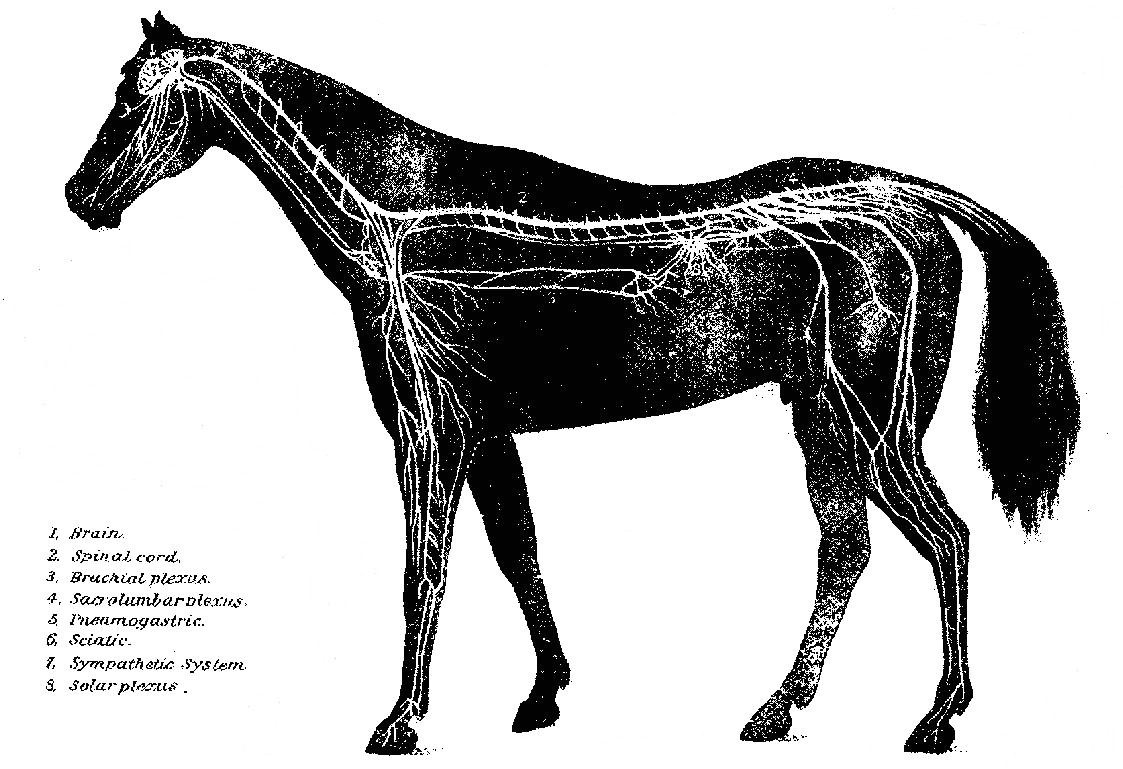

PLATE XIX.
THE NERVOUS SYSTEM.

If this treatment fails to give relief, the disease will pass into the advanced stages, or, if the animal has been neglected in the early stages, the treatment must be supplanted with the hypodermic injection of ergotin, in 5-grain doses, dissolved in 1 dram of water, every six hours. The limbs may be poulticed above the fetlocks with mustard. Warm blanketing, to promote perspiration, is to be observed always when there is no excessive perspiration.

If the disease becomes chronic (encephalitis or meningitis), we must place our reliance upon alteratives and tonics, with such incidental treatment as special symptoms may demand. Iodid of potassium in 2-dram doses should be given three times a day and 1 dram of calomel once a day to induce absorption of effusions or thickened membranes. Tonics, in the form of iodid of iron in 1-dram doses, to which is added 2 drams of powdered hydrastis, may also be given every six or eight hours, as soon as the active fever has abated. After the disappearance of the acute symptoms, blisters (cantharides ointment) may be applied behind the poll. When paralytic effects remain after the disappearance of all other symptoms, sulphate of strychnia in 2-grain doses, in combination with the other tonics, may be given twice a day and be continued until it produces muscular twitching. In some cases of paralysis, as of the lips or throat, benefit may be derived from the moderate use of the electric battery. Many of the recoveries will, however, under the most active and early treatment, be but partial, and in all cases the animals become predisposed to subsequent attacks. A long time should be allowed to pass before the animal is exposed to severe work or great heat. When the disease depends upon mechanical injuries, they have to be treated and all causes of irritation to the brain removed. If it is due to stable miasma, uremic poisoning, pyemia, influenza, rheumatism, toxic agents, etc., they should receive prompt attention for their removal or mitigation.

Cerebral softening, abscess, and sclerosis are practically inaccessible to treatment, otherwise than such relief as may be afforded by the administration of opiates and general tonics, and, in fact, the diagnosis is largely presumptive.

terça-feira, 3 de novembro de 2015

With its inflammatory products and exudations

With its inflammatory products and exudations

With its inflammatory products and exudations


Treatment. In all acute attacks of inflammation involving the membranes or cerebral masses, it is the pressure from the distended and engorged blood vessels and the rapid accumulation of inflammatory products that endangers the life of the animal in even the very early stage of the disease. The earlier the treatment is commenced to lessen the danger of fatal pressure from the engorged blood vessels, the less effusion and smaller number of inflammatory products we have to contend with later. The leading object, then, to be accomplished in the treatment of the first stage of encephalitis, meningitis, or cerebritis, and before a dangerous degree of effusion or exudation has taken place, is to relieve the engorgement of the blood vessels and thereby lessen the irritation or excitability of the affected structures. If the attempt to relieve the engorgement in the first stage has been only partially successful, and the second stage, with its inflammatory products and exudations, whether serous or plastic, has set in, then the main objects in further treatment are to keep up the strength of the animal and hasten the absorption of the exudative products as much as possible. To obtain these results, when the animal is found in the initial stage of the disease, if there is unnatural excitability or stupor with increase of temperature and quickened pulse, we should apply cold to the head in the form of cold water or ice. For this purpose cloths or bags may be used, and they should be renewed as often as necessary. If the disease is still in its early stages and the animal is strong, bleeding from the jugular vein may be beneficial. Good results are to be expected only during the stage of excitement, while there is a strong, full pulse and the mucous membranes of the head are red from a plentiful supply of blood. The finger should be kept on the pulse and the blood allowed to flow until there is distinct softening of the pulse. As soon as the animal recovers somewhat from the shock of the bleeding the following medicine should be made into a ball or dissolved in a pint of warm water and be given at one dose: Barbados aloes, 7 drams; calomel, 2 drams; powdered ginger, 1 dram; tincture of aconite, 20 drops.

segunda-feira, 2 de novembro de 2015

Or inflammation of the interior of the brain

Or inflammation of the interior of the brain

Or inflammation of the interior of the brain


In cerebritis, or inflammation of the interior of the brain, there is a tendency to softening and suppuration and the formation of abscesses. In some cases the abscesses are small and numerous, surrounded with a softened condition of the brain matter, and sometimes we may find one large abscess. In cases of recent development the walls of the abscesses are fringed and ragged and have no lining membrane. In older or chronic cases the walls of the abscesses are generally lined with a strong membrane, often having the appearance of a sac or cyst, and the contents have a very offensive odor.

domingo, 1 de novembro de 2015

In addition to the serous effusion

In addition to the serous effusion

In addition to the serous effusion


In meningitis, especially in chronic cases, in addition to the serous effusion, there are changes which may be regarded as characteristic in the formation of a delicate and highly vascular layer or layers of membrane or organized structure on the surface of the dura mater, and also indications of hemorrhages in connection with the membranous formations. Hematoma, or blood tumors, may be found embedded in this membrane. In some cases the hemorrhages are copious, causing paralysis or apoplexy, followed by speedy death. The meningitis may be suppurative. In this case a puslike exudate is found between the membranes covering the brain.

sábado, 31 de outubro de 2015

If death has occurred at a later period of the disease

If death has occurred at a later period of the disease

If death has occurred at a later period of the disease


Lesions. On making post-mortem examinations of horses which have died in the first stages of either of these diseases we find an excessive engorgement of the capillaries and small blood vessels, with correspondingly increased redness and changes in both the contents and the walls of the vessels. If death has occurred at a later period of the disease, it will be found that, in addition to the redness and engorgement, an exudation of the contents of the blood vessels into the tissues and upon the surfaces of the inflamed parts has supervened. If the case has been one of encephalitis, there will usually be found more or less watery fluid in the ventricles (natural cavities in the brain), in the subarachnoid space, and a serous exudation between the convolutions and interstitial spaces of the gray matter under the membranes of the brain. The quantity of fluid varies in different cases. Exudations of a membranous character may be present, and are found attached to the surfaces of the pia mater.

sexta-feira, 30 de outubro de 2015

Causing their ultimate disappearance

Causing their ultimate disappearance

Causing their ultimate disappearance


Cerebral sclerosis sometimes follows inflammation in the structure of the brain affecting the connective tissues, which eventually become hypertrophied and press upon nerve cells and fibers, causing their ultimate disappearance, leaving the parts hard and indurated. This condition gives rise to a progressive paralysis and may extend along a certain bundle of fibers into the spinal cord. Complete paralysis almost invariably supervenes and causes death.

quinta-feira, 29 de outubro de 2015

Softening and abscess of the brain are terminations of cerebritis

Softening and abscess of the brain are terminations of cerebritis

Softening and abscess of the brain are terminations of cerebritis


Softening and abscess of the brain are terminations of cerebritis. It may also be due to an insufficient supply of blood as a result of diseased cerebral arteries and of apoplexy. The symptoms are drowsiness, vertigo, or attacks of giddiness, increased timidity, or fear of familiar objects, paralysis of one limb, hemiplegia, imperfect control of the limbs, and usually a weak, intermittent pulse. In some cases the symptoms are analogous to those of apoplexy. The character of the symptoms depends upon the seat of the softening or abscess within the brain.

quarta-feira, 28 de outubro de 2015

Following these symptoms there are depression

Following these symptoms there are depression

Following these symptoms there are depression


Following these symptoms there are depression, loss of power and consciousness, lack of ability or desire to move, and usually fall of temperature. At this stage the horse stands with legs propped, the head hanging or resting on the manger, the eyes partly closed, and does not respond when spoken to or when struck with a whip.

Chronic encephalitis or meningitis may succeed the acute stage, or may be due to stable miasma, blood poison, narcotism, lead poisoning, etc. This form may not be characterized in its initial stages by excitability, quick and hard pulse, and high fever. The animal usually appears at first stupid; eats slowly; the pupil of the eye does not respond to light quickly; the animal often throws his head up or shakes it as if suffering sudden twinges of pain. He is slow and sluggish in his movements, or there may be partial paralysis of one limb, one side of the face, neck, or body. These symptoms, with some variations, may be present for several days and then subside, or the disease may pass into the acute stage and terminate fatally. Chronic encephalitis may effect an animal for ten days or two weeks without much variation in the symptoms before the crisis is reached. If improvement commences, the symptoms usually disappear in the reverse order to that in which they developed, with the exception of the paralytic effects, which remain intractable or permanent. Paralysis of certain sets of muscles is a very common result of chronic, subacute, and acute encephalitis, and is due to softening of the brain or to exudation into the cavities of the brain or arachnoid space.

terça-feira, 27 de outubro de 2015

Usually the first symptoms indicate mental excitement

Usually the first symptoms indicate mental excitement

Usually the first symptoms indicate mental excitement


Symptoms. The diseases here grouped together are accompanied with a variety of symptoms, almost none of which, however, are associated so definitely with a special pathological process as to point unmistakably to a given lesion. Usually the first symptoms indicate mental excitement, and are followed by symptoms indicating depression. Acute encephalitis may be ushered in by an increased sensibility to noises, with more or less nervous excitability, contraction of the pupils of the eyes, and a quick, hard pulse. In very acute attacks these symptoms, however, are not always noted. This condition will soon be followed by muscular twitchings, convulsive or spasmodic movements, eyes wide open with shortness of sight. The animal becomes afraid to have his head handled. Convulsions and delirium will develop, with inability of muscular control, or stupor and coma may supervene. When the membranes are greatly implicated, convulsions and delirium with violence may be expected, but if the brain substances are principally affected stupor and coma will be the prominent symptoms. In the former condition the pulse will be quick and hard; in the latter, soft and depressed, with often a dilatation of the pupils, and deep, slow, stertorous breathing. The symptoms may follow one another in rapid succession, and the disease approach a fatal termination within 12 hours. In subacute attacks the symptoms are better defined, and the animal seldom dies before the third day. Within three or four days gradual improvement may become manifest, or cerebral softening with partial paralysis may occur. In all cases of encephalitis there is a marked rise in temperature from the very onset of the disease, with a tendency to increase until the most alarming symptoms develop, succeeded by a decrease when coma becomes manifest. The violence and character of the symptoms greatly depend upon the extent and location of the structures involved. Thus, in some cases there may be marked paralysis of certain muscles, while in others there may be spasmodic rigidity of muscles in a certain region. Very rarely the animal becomes extremely violent early in the attack, and by rearing up, striking with the fore feet, or falling over, may do himself great injury. Usually, however, the animal maintains the standing position, propping himself against the manger or wall, until he falls from inability of muscular control, or from unconsciousness. Occasionally, in his delirium, he may go through a series of automatic movements, such as trotting or walking, and, if loose in a stall, will move around persistently in a circle. Early and persistent constipation of the bowels is a marked symptom in nearly all acute affections of the brain; retention of the urine, also, is frequently observed.

segunda-feira, 26 de outubro de 2015

Exposure to extreme heat or cold

Exposure to extreme heat or cold

Exposure to extreme heat or cold


Causes. Exposure to extreme heat or cold, sudden and extreme changes of temperature, excessive continued cerebral excitement, too much nitrogenous feed, direct injuries to the brain, such as concussion, or from fracture of the cranium, overexertion, sometimes as sequelae to influenza, pyemia, poisons having a direct influence upon the encephalic mass, extension of inflammation from neighboring structures, food poisoning, tumors, parasites, metastatic abscesses, etc.

domingo, 25 de outubro de 2015

Inflammation of The Brain And Its Membranes (Encephalitis, Meningitis, Cerebritis).

Inflammation of The Brain And Its Membranes (Encephalitis, Meningitis, Cerebritis).

Inflammation of The Brain And Its Membranes (Encephalitis, Meningitis, Cerebritis).


Inflammation may attack these membranes singly, or any one of the anatomical divisions of the nerve matter, or it may invade the whole at once. Practical experience, however, teaches us that primary inflammation of the dura mater is of rare occurrence, except in direct mechanical injuries to the head or diseases of the bones of the cranium. Neither is the arachnoid often affected with acute inflammation, except as a secondary result. The pia mater is most commonly the seat of inflammation, acute and subacute, but from its intimate relation with the surface of the brain the latter very soon becomes involved in the morbid changes. Practically, we can not separate inflammation of the pia mater from that of the brain proper. Inflammation may, however, exist in the center of the great nerve masses the cerebrum, cerebellum, pons Varolii, or medulla at the base of the brain without involving the surface. When, therefore, inflammation invades the brain and its enveloping membranes it is properly called encephalitis; when the membranes alone are affected it is called meningitis, or the brain substance alone cerebritis. Since all the conditions merge into one another and can scarcely be recognized separately during the life of the animal, they may here be considered together.

sábado, 24 de outubro de 2015

Forty-two or forty-three in number

Forty-two or forty-three in number

Forty-two or forty-three in number


The spinal nerves, forty-two or forty-three in number, arise each by two roots, a superior or sensory, and an inferior or motor. The nerves originating from the brain are twenty-four in number, and arranged in pairs, which are named first, second, third, etc., counting from before backward. They also receive special names, according to their functions or the parts to which they are distributed, viz:

1. Olfactory.
2. Optic.
3. Oculo-motor.
4. Pathetic.
5. Trifacial.
6. Abducens.
7. Facial.
8. Auditory.
9. Glossopharyngeal.
10. Pneumogastric.
11. Spinal accessory.
12. Hypoglossal.

sexta-feira, 23 de outubro de 2015

It is an irregularly cylindrical structure

It is an irregularly cylindrical structure

It is an irregularly cylindrical structure


The spinal cord, or spinal marrow, is that part of the cerebrospinal system which is contained in the spinal canal of the backbone, and extends from the medulla oblongata to a short distance behind the loins. It is an irregularly cylindrical structure, divided into two lateral, symmetrical halves by fissures. The spinal cord terminates posteriorly in a pointed extremity, which is continued by a mass of nerve trunks cauda equinae. A transverse section of the cord reveals that it is composed of white matter externally and of gray matter internally. The spinal cord does not fill the whole spinal canal. The latter contains, besides, a large venous sinus, fatty matter, the membranes of the cord, and the cerebrospinal fluid.

quinta-feira, 22 de outubro de 2015

Occupies the anterior portion of the cranial cavity

Occupies the anterior portion of the cranial cavity

Occupies the anterior portion of the cranial cavity


The cerebrum, or brain proper, occupies the anterior portion of the cranial cavity. It is ovoid in shape, with an irregular, flattened base, and consists of lateral halves or hemispheres. The greater part of the cerebrum is composed of white matter. The hemispheres of the cerebrum are usually said to be the seat of all psychical activities. Only when they are intact are the process of feeling, thinking, and willing possible. After they are destroyed the organism comes to be like a complicated machine, and its activity is only the expression of the internal and external stimuli which act upon it.

quarta-feira, 21 de outubro de 2015

Immediately above the medulla oblongata

Immediately above the medulla oblongata

Immediately above the medulla oblongata


The cerebellum is lodged in the posterior part of the cranial cavity, immediately above the medulla oblongata; it is globular or elliptical in shape, the transverse diameter being greatest. The body of the cerebellum is composed of gray matter externally and of white matter in the center. The cerebellum has the function of co-ordinating movements; that is, of so associating them as to cause them to accomplish a definite purpose. Injuries to the cerebellum cause disturbances of the equilibrium but do not interfere with the will power or intelligence.

terça-feira, 20 de outubro de 2015

The medulla oblongata is the prolongation of the spinal cord

The medulla oblongata is the prolongation of the spinal cord

The medulla oblongata is the prolongation of the spinal cord


The medulla oblongata is the prolongation of the spinal cord, extending to the pons Varolii. This portion of the brain is very large in the horse: it is pyramidal in shape, the narrowest part joining the cord.

The pons Varolii is the transverse projection on the base of the brain, between the medulla oblongata and the peduncles of the cerebrum.

segunda-feira, 19 de outubro de 2015

Which may be divided into four parts the medulla oblongata

Which may be divided into four parts the medulla oblongata

Which may be divided into four parts the medulla oblongata


The brain is that portion of the cerebrospinal axis within the cranium, which may be divided into four parts the medulla oblongata, the cerebellum, the pons Varolii, and the cerebrum and it is covered by three membranes, called the meninges. The outer of these membranes, the dura mater, is a thick, white, fibrous membrane which lines the cavity of the cranium, forming the internal periosteum of the bones; it is continuous with the spinal cord to the extremity of the canal. The second, the arachnoid, is a delicate serous membrane, and loosely envelops the brain and spinal cord; it forms two layers, having between them the arachnoid space which contains the cerebrospinal fluid, the use of which is to protect the spinal cord and brain from pressure. The third, or inner, the pia mater, is closely adherent to the entire surface of the brain, but is much thinner and more vascular than when it reaches the spinal cord, which it also envelops, and is continued to form the sheaths of the spinal nerves.

domingo, 18 de outubro de 2015

Nerves which convey impressions to the centers are termed sensory

Nerves which convey impressions to the centers are termed sensory

Nerves which convey impressions to the centers are termed sensory


Nerves which convey impressions to the centers are termed sensory, or centripetal, and those which transmit stimulus from the centers to organs of motion are termed motor, or centrifugal. The function of the nervous system may, therefore, be defined in the simplest terms, as follows: It is intended to associate the different parts of the body in such a manner that stimulus applied to one organ may excite or depress the activity of another.

sábado, 17 de outubro de 2015

Nerves are thrown into a state of excitement when stimulated

Nerves are thrown into a state of excitement when stimulated

Nerves are thrown into a state of excitement when stimulated


Nerves are thrown into a state of excitement when stimulated, and are, therefore, said to possess excitable or irritable properties. The stimuli may be applied to, or may act upon, any part of the nerve. Nerves may be paralyzed by continuous pressure being applied. When the nerves divide into branches, there is never any splitting up of their ultimate fibers, nor yet is there ever any coalescing of them; they retain their individuality from their source to their termination.

sexta-feira, 16 de outubro de 2015

Act as physiological centers for automatic

Act as physiological centers for automatic

Act as physiological centers for automatic


The nerve fibers represent a conducting apparatus and serve to place the central nervous organs in connection with peripheral end organs. The nerve cells, however, besides transmitting impulses, act as physiological centers for automatic, or reflex, movements, and also for the sensory, perceptive, trophic, and secretory functions. A nerve consists of a bundle of tubular fibers, held together by a dense areolar tissue, and inclosed in a membranous sheath the neurilemma. Nerve fibers possess no elasticity, but are very strong. Divided nerves do not retract.

quinta-feira, 15 de outubro de 2015

Distinguishable by their color

Distinguishable by their color

Distinguishable by their color


Two substances, distinguishable by their color, namely, the white or medullary and the gray or cortical substance, enter into the formation of nervous matter. Both are soft, fragile, and easily injured, in consequence of which the principal nervous centers are well protected by bony coverings. The nervous substances present two distinct forms nerve fibers and nerve cells. An aggregation of nerve cells constitutes a nerve ganglion.

quarta-feira, 14 de outubro de 2015

The central organ consists of a chain of ganglia

The central organ consists of a chain of ganglia

The central organ consists of a chain of ganglia


In the second, the central organ consists of a chain of ganglia, connected by nerve cords, which extends on each side of the spine from the head to the rump. The nerves of this system are distributed to the involuntary muscles, mucous membrane, viscera, and blood vessels.

The two systems have free intercommunication, ganglia being at the junctions.

terça-feira, 13 de outubro de 2015

The other elongated spinal cord

The other elongated spinal cord

The other elongated spinal cord


In the first, the center is made up of two portions one large and expanded (the brain) placed in the cranial cavity; the other elongated (spinal cord), continuous with the brain, and lodged in the canal of the vertebral column. The peripheral portion of this system consists of the cerebrospinal nerves, which leave the axis in symmetrical pairs and are distributed to the skin, the voluntary muscles, and the organs.

segunda-feira, 12 de outubro de 2015

(Pl. Xix.)

(Pl. Xix.)

(Pl. Xix.)


The nervous system may be regarded as consisting of two sets of organs, peripheral and central, the function of one being to establish a communication between the centers and the different parts of the body, and that of the other to generate nervous force. The whole may be arranged under two divisions: First, the cerebrospinal system; second, the sympathetic or ganglionic system. Each is possessed of its own central and peripheral organs.

domingo, 11 de outubro de 2015

Sore Teats, Scabs, Cracks, Warts.

Sore Teats, Scabs, Cracks, Warts.

Sore Teats, Scabs, Cracks, Warts.


By the act of sucking, especially in cold weather, the teats are subject to abrasions, cracks, and scabs, and as the result of such irritation, or independently, warts sometimes grow and prove troublesome. The warts should be clipped off with sharp scissors and their roots burned with a solid pencil of lunar caustic. This is best done before parturition to secure healing before suckling begins. For sore teats use an ointment of vaseline 1 ounce, balsam of tolu 5 grains, and sulphate of zinc 5 grains.


sábado, 10 de outubro de 2015

Tumors of The Udder.

Tumors of The Udder.

Tumors of The Udder.


As the result of inflammation of the udder it may become the seat of an indurated diseased growth, which may go on growing and seriously interfere with the movement of the hind limbs. If such swellings do not give way in their early stages to treatment by iodin, the only resort is to cut them out with a knife. As the gland is often implicated and has to be removed, such mares can not in the future suckle their colts and therefore should not be bred.

sexta-feira, 9 de outubro de 2015

The treatment is simple so long as there is only congestion

The treatment is simple so long as there is only congestion

The treatment is simple so long as there is only congestion


Treatment. The treatment is simple so long as there is only congestion. Active rubbing with lard or oil, or, better, camphorated oil, and the frequent drawing off of the milk, by the foal or with the hand, will usually bring about a rapid improvement. When active inflammation is present, fomentation with warm water may be kept up for an hour and followed by the application of the camphorated oil, to which has been added some carbonate of soda and extract of belladonna. A dose of laxative medicine (4 drams Barbados aloes) will be of service in reducing fever, and one-half ounce saltpeter daily will serve a similar end. In case the milk coagulates in the udder and can not be withdrawn, or when the liquid becomes fetid, a solution of 20 grains carbonate of soda and 10 drops carbolic acid dissolved in an ounce of water should be injected into the teat. In doing this it must be noted that the mare has three separate ducts opening on the summit of each teat and each must be carefully injected. To draw off the fetid product it may be needful to use a small milking tube, or spring teat dilator designed by the writer. (Plate XIV, figs. 2 and 3.) When pus forms and points externally and can not find a free escape by the teat, the spot where it fluctuates must be opened freely with the knife and the cavity injected daily with the carbolic-acid lotion. When the gland becomes hard and indolent, it may be rubbed daily with iodin ointment 1 part, vaseline 6 parts.

quinta-feira, 8 de outubro de 2015

Diseases of The Udder And Teats. Congestion And Inflammation of The Udder.

Diseases of The Udder And Teats. Congestion And Inflammation of The Udder.

Diseases of The Udder And Teats. Congestion And Inflammation of The Udder.


This is comparatively rare in the mare, though in some cases the udder becomes painfully engorged before parturition, and a doughy swelling, pitting on pressure, extends forward on the lower surface of the abdomen. When this goes on to active inflammation, one or both of the glands becomes enlarged, hot, tense, and painful; the milk is dried up or replaced by a watery or reddish, serous fluid, which at times becomes fetid; the animal walks lame, loses appetite, and shows general disorder and fever. The condition may end in recovery, in abscess, induration, or gangrene, and, in some cases, may lay the foundation for a tumor of the gland.

quarta-feira, 7 de outubro de 2015

Leucorrhea.

Leucorrhea.

Leucorrhea.


This is a white, glutinous, chronic discharge, the result of a continued, subacute inflammation of the mucous membrane of the womb. Like the discharge of acute inflammation, it contains many forms of bacteria, by some of which it is manifestly inoculable on the penis of the stallion, producing ulcers and a specific, gonorrheal discharge.

Treatment may consist in the internal use of tonics (sulphate of iron, 3 drams, daily) and the washing out of the womb, as described under the last heading, followed by an astringent antiseptic injection (carbolic acid, 2 teaspoonfuls; tannic acid, 1/2 dram; water, 1 quart). This may be given two or three times a day.

terça-feira, 6 de outubro de 2015

And into which a funnel is fitted

And into which a funnel is fitted

And into which a funnel is fitted


Treatment. In treatment the first thing to be sought is the removal of all offensive and irritant matters from the womb through a caoutchouc tube introduced into the womb, and into which a funnel is fitted. Warm water should be passed until it comes away clear. To insure that all the womb has been washed out, the oiled hand may be introduced to carry the end of the tube into the two horns successively. When the offensive contents have been thus removed, the womb should be injected with a quart of water holding in solution 1 dram permanganate of potash, or, in the absence of the latter, 2 teaspoonfuls of carbolic acid, twice daily. Fomentation of the abdomen, or the application of a warm flaxseed poultice, may greatly relieve. Acetanilid, in doses of half an ounce, twice or thrice a day, or sulphate of quinia in doses of one-third ounce, may be employed to reduce the fever. If the great prostration indicates septic poisoning, large doses (one-half ounce) bisulphite of soda, or salicylate of soda, or sulphate of quinin may be resorted to.

segunda-feira, 5 de outubro de 2015

The symptoms are ushered in by shivering

The symptoms are ushered in by shivering

The symptoms are ushered in by shivering


Symptoms. The symptoms are ushered in by shivering, staring coat, small, rapid pulse, elevated temperature, accelerated breathing, loss of appetite, with arched back, stiff movement of the body, looking back at the flanks, and uneasy motions of the hind limbs, discharge from the vulva of a liquid at first watery, reddish, or yellowish, and later it may be whitish or glairy, and fetid or not in different cases. Tenderness of the abdomen shown on pressure is especially characteristic of cases affecting the peritoneum or lining of the belly, and is more marked lower down. If the animal survives, the inflammation tends to become chronic and attended by a whitish mucopurulent discharge. If, on the contrary, it proves fatal, death is preceded by extreme prostration and weakness from the general septic poisoning.

domingo, 4 de outubro de 2015

Inflammation of The Womb And Peritoneum.

Inflammation of The Womb And Peritoneum.

Inflammation of The Womb And Peritoneum.


These may result from injuries sustained by the womb during or after parturition, from exposure to cold or wet, or from the irritant infective action of putrid products within the womb. Under the inflammation the womb remains dilated and flaccid, and decomposition of its secretions almost always occurs, so that the inflammation tends to assume a putrid character and general septic infection is likely to occur.

sábado, 3 de outubro de 2015

Laminitis, or Founder, Following Parturition.

Laminitis, or Founder, Following Parturition.

Laminitis, or Founder, Following Parturition.


This sometimes follows on inflammation of the womb, as it frequently does on disorder of the stomach. Its symptoms agree with those of the common form of founder, and treatment need not differ.

sexta-feira, 2 de outubro de 2015

Blood Clots in The Walls of The Vagina.

Blood Clots in The Walls of The Vagina.

Blood Clots in The Walls of The Vagina.


(See "Effusion of blood in the vaginal walls," p. 190.)

quinta-feira, 1 de outubro de 2015

Ruptures of The Vagina.

Ruptures of The Vagina.

Ruptures of The Vagina.


These are attended with dangers similar to those belonging to rupture of the womb, and in addition by the risk of protrusion of the bladder, which appears through the lips of the vulva as a red, pyriform mass. Sometimes such lacerations extend downward into the bladder, and in others upward into the terminal gut (rectum). In still other cases the anus is torn so that it forms one common orifice with the vulva.

Too often such cases prove fatal, or at least a recovery is not attained, and urine or feces or both escape freely into the vagina. The simple laceration of the anus is easily sewed up, but the ends of the muscular fibers do not reunite and the control over the lower bowel is never fully reacquired. The successful stitching up of the wound communicating with the bladder or the rectum requires unusual skill and care, and though I have succeeded in a case of the latter kind, I can not advise the attempt by unprofessional persons.

quarta-feira, 30 de setembro de 2015

Rupture, or Laceration, of The Womb.

Rupture, or Laceration, of The Womb.

Rupture, or Laceration, of The Womb.


This may occur from the feet of the foal during parturition, or from ill-directed efforts to assist, but it is especially liable to take place in the everted, congested, and friable organ. The resultant dangers are bleeding from the wound, escape of the bowels through the opening and their fatal injury by the mare's feet or otherwise, and peritonitis from the extension of inflammation from the wound and from the poisonous action of the septic liquids of the womb escaping into the abdominal cavity. The first object is to close the wound, but unless in eversion of the womb this is practically impossible. In the last-named condition the wound must be carefully and accurately sewed up before the womb is returned. After its return, the womb must be injected daily with an antiseptic solution (borax, one-half ounce, or carbolic acid, 3 drams to a quart of tepid water). If inflammation threatens, the abdomen may be bathed continuously with hot water by means of a heavy woolen rag, and large doses of opium (one-half dram) may be given twice or thrice daily.

terça-feira, 29 de setembro de 2015

Eversion of The Womb.

Eversion of The Womb.

Eversion of The Womb.


If the womb fails to contract after difficult parturition, the after-pains will sometimes lead to the fundus passing into the body of the organ and passing through that and the vagina until the whole inverted organ appears externally and hangs down on the thighs. The result is rapid engorgement and swelling of the organ, impaction of the rectum with feces, and distention of the bladder with urine, all of which conditions seriously interfere with the return of the mass. In returning the womb the standing is preferable to the recumbent position, as the abdomen is more pendent and there is less obstruction to the return. It may, however, be necessary to put hobbles on the hind limbs to prevent the mare from kicking. A clean sheet should be held beneath the womb, and all filth, straw, and foreign bodies washed from its surface. Then with a broad, elastic (india-rubber) band, or in default of that a long strip of calico 4 or 5 inches wide, wind the womb as tightly as possible, beginning at its most dependent part (the extremity of the horn). This serves two good ends. It squeezes out into the general circulation the enormous mass of blood which engorged and enlarged the organ, and it furnishes a strong protective covering for the now delicate, friable organ, through which it may be safely manipulated without danger of laceration. The next step may be the pressure on the general mass while those portions next the vulva are gradually pushed in with the hands; or the extreme lowest point (the end of the horn) may be turned within itself and pushed forward into the vagina by the closed fist, the return being assisted by manipulations by the other hand, and even by those of assistants. By either mode the manipulations may be made with almost perfect safety so long as the organ is closely wrapped in the bandage. Once a portion has been introduced into the vagina the rest will usually follow with increasing ease, and the operation should be completed with the hand and arm extended the full length within the womb and moved from point to point so as to straighten out all parts of the organ and insure that no portion still remain inverted within another portion. Should any such partial inversion be left it will give rise to straining, under the force of which it will gradually increase until the whole mass will be protruded as before. The next step is to apply a truss as an effectual mechanical barrier to further escape of the womb through the vulva. The simplest is made with two 1-inch ropes, each about 18 feet long, each doubled and interwoven at the bend, as seen in Plate XIV, figure 4. The ring formed by the interlacing of the two ropes is adjusted around the vulva, the two ends of the one rope are carried up on the right and left of the tail and along the spine, being wound around each other in their course, and are finally tied to the upper part of the collar encircling the neck. The remaining two ends, belonging to the other rope, are carried downward and forward between the thighs and thence forward and upward on the sides of the belly and chest to be attached to the right and left sides of the collar. These ropes are drawn tightly enough to keep closely applied to the opening without chafing, and will fit still more securely when the mare raises her back to strain. It is desirable to tie the mare short so that she may be unable to lie down for a day or two, and she should be kept in a stall with the hind parts higher than the fore. Violent straining may be checked by full doses of opium (one-half dram), and any costiveness or diarrhea should be obviated by a suitable laxative or binding diet.

In some mares the contractions are too violent to allow of the return of the womb, and full doses of opium one-half dram, laudanum 2 ounces, or chloral hydrate 1 ounce, may be demanded, or the mare must be rendered insensible by ether or chloroform.

segunda-feira, 28 de setembro de 2015

Flooding, or Bleeding From The Womb.

Flooding, or Bleeding From The Womb.

Flooding, or Bleeding From The Womb.


This is rare in the mare, but not unknown, in connection with a failure of the womb to contract on itself after parturition, or with eversion of the womb (casting the withers), and congestion or laceration. If the blood accumulates in the flaccid womb, the condition may be suspected only by reason of the rapidly advancing weakness, swaying, unsteady gait, hanging head, paleness of the eyes and other mucous membranes, and weak, small, failing pulse. The hand introduced into the womb detects the presence of the blood partly clotted. If the blood escapes by the vulva, the condition is evident.

Treatment consists in evacuating the womb of its blood clots, giving a large dose of powdered ergot of rye, and in the application of cold water or ice to the loins and external generative organs. Besides this, a sponge impregnated with a strong solution of alum, or, still better, with tincture of muriate of iron, may be introduced into the womb and squeezed so as to bring the liquid in contact with the walls generally.

domingo, 27 de setembro de 2015

Removal of the abdominal viscera

Removal of the abdominal viscera

Removal of the abdominal viscera


Removal of the abdominal viscera. In case the belly is unduly large, from decomposition, tumors, or otherwise, it may be needful to lay it open with the knife and cut or tear out the contents.

Removal of the thoracic viscera. To diminish the bulk of the chest it has been found advisable to cut out the breastbone, remove the heart and lungs, and allow the ribs to collapse with the lower free ends overlapping each other.

Dissection of the trunk. In case it becomes necessary to remove other portions of the trunk, we should follow the general rule of preserving the skin so that all manipulations can be made inside this as a protector, that it may remain available as a means of exercising traction on the remaining part of the body, and as a covering to protect the vaginal walls against injuries from bones while such part is passing.

sábado, 26 de setembro de 2015

Reuff cuts his way into the pelvis of the foal

Reuff cuts his way into the pelvis of the foal

Reuff cuts his way into the pelvis of the foal


Reuff cuts his way into the pelvis of the foal, and with a knife separates the pelvic bones from the loins, then skinning the quarter draws out these pelvic bones by means of ropes and hooks, and along with them the hind limbs.

The hind limbs having been removed by one or the other of these procedures, the loose skin detached from the pelvis is used as a means of traction and delivery is effected. In case of a monstrosity with extra hind limbs, it may be possible to bring these up into the passage and utilize them for traction.

sexta-feira, 25 de setembro de 2015

Then with cords round the pelvic bones

Then with cords round the pelvic bones

Then with cords round the pelvic bones


Cartwright recommends to make free incisions round the hip joints and tear through the muscles when they can not be cut; then with cords round the pelvic bones, and hooks inserted in the openings in the floor of the pelvis to drag out the pelvic bones; then put cords around the heads of the thigh bones and extract them; then remove the intestines; finally, by means of the loose, detached skin, draw out the body with the remainder of the hind limbs bent forward beneath it.

quinta-feira, 24 de setembro de 2015

In case the limb is turned forward from the hip

In case the limb is turned forward from the hip

In case the limb is turned forward from the hip


In case the limb is turned forward from the hip, and the fetus so wedged into the passage that turning is impossible, the case is very difficult. I have repeatedly succeeded by cutting in on the hip joint and disarticulating it, then dissecting the muscles back from the upper end of the thigh bone. A noose was placed around the neck of the bone and pulled on forcibly, while any unduly resisting structures were cut with the knife.

quarta-feira, 23 de setembro de 2015

In case the limb is bent forward at the hock

In case the limb is bent forward at the hock

In case the limb is bent forward at the hock


In case the limb is bent forward at the hock, a rope is passed round that and pulled so as to bring the point of the hock between the lips of the vulva. The hamstring and the lateral ligaments of the hock are now cut through, and the limbs extended by a rope tied round the lower end of the long bone above (tibia). In case it is still needful to remove the upper part of the limb, the further procedure is the same as described in the last paragraph.

terça-feira, 22 de setembro de 2015

An embryotomy knife is desirable

An embryotomy knife is desirable

An embryotomy knife is desirable


Amputation of the fore limbs. This may usually be begun on the fetlock of the limb projecting from the vulva. An embryotomy knife is desirable. This knife consists of a blade with a sharp, slightly hooked point, and one or two rings in the back of the blade large enough to fit on the middle finger, while the blade is protected in the palm of the hand. (See Plate XIII, fig. 4.) Another form has the blade inserted in a mortise in the handle, from which it is pushed out by a movable button when wanted. First place a noose around the fetlock of the limb to be amputated, cut the skin circularly entirely around the fetlock, then make an incision on the inner side of the limb from the fetlock up to the breastbone. Next dissect the skin from the limb, from the fetlock up to the breastbone on the inner side, and as far up on the shoulder blade as possible on the outer side. Finally, cut through the muscles attaching the limb to the breastbone, and employ strong traction on the limb, so as to drag out the whole limb, shoulder blade included. The muscles around the upper part of the shoulder blade are easily torn through and need not be cut, even if that were possible. In no case should the fore limb be removed unless the shoulder blade is taken with it, as that furnishes the greatest obstruction to delivery, above all when it is no longer advanced by the extension of the fore limb, but is pressed back so as to increase the already thickest posterior portion of the chest. The preservation of the skin from the whole limb is advantageous in various ways; it is easier to cut it circularly at the fetlock than at the shoulder; it covers the hand and knife in making the needful incisions, thus acting as a protection to the womb; and it affords a means of traction on the body after the limb has been removed. In dissecting the skin from the limb the knife is not needful at all points; much of it may be stripped off with the fingers or knuckles, or by a blunt, iron spud, pushed up inside the hide, which is meanwhile held tense to render the spud effective.

segunda-feira, 21 de setembro de 2015

But requires more anatomical knowledge

But requires more anatomical knowledge

But requires more anatomical knowledge


Amputation of the hind limb. This may be required when there are extra hind limbs or when the hind limbs are bent forward at hock or hip joint. In the former condition the procedure resembles that for removal of a fore limb, but requires more anatomical knowledge. Having noosed the pastern, a circular incision is made through the skin around the fetlock, and a longitudinal one from that up to the groin, and the skin is dissected from the limb as high up as can be reached, over the croup, if possible. Then cut through the muscles around the hip joint, and, if possible, the two interarticular ligaments of the joint (pubofemoral and round), and extract the limb by strong dragging.

domingo, 20 de setembro de 2015

The head is secured by a hook in the lower jaw

The head is secured by a hook in the lower jaw

The head is secured by a hook in the lower jaw


Amputation of the head. This is easy when both fore limbs are turned back and the head alone has made its exit in part. It is more difficult when the head is still retained in the passages or womb, as in double-headed monsters. The head is secured by a hook in the lower jaw, or in the orbit, or by a halter, and the skin is divided circularly around the lower part of the face or at the front of the ears, according to the amount of head protruding. Then an incision is made backward along the line of the throat, and the skin dissected from the neck as far back as possible. Then the muscles and other soft parts of the neck are cut across, and the bodies of two vertebra (neck bones) are severed by cutting completely across the cartilage of the joint. The bulging of the ends of the bones will serve to indicate the seat of the joint. The head and detached portion of the neck may now be removed by steady pulling. If there is still an obstacle, the knife may be again used to sever any obstinate connections. In the case of a double-headed monster, the whole of the second neck must be removed with the head. When the head has been detached, a rope should be passed through the eyeholes, or through an artificial opening in the skin, and tied firmly around the skin, to be employed as a means of traction when the missing limbs or the second head have been brought up into position.